
Protein Labeling Reagents


Cayman Chemical Fluorescein 5-isothiocyanate
An amine- and thiol-reactive fluorescent probe; forms conjugates with proteins, lipids, and other molecules for detection by a variety of fluorescent-based applications; ex/em = 494/519 nm, respectively

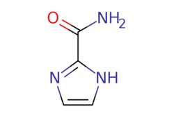
eMolecules 16093-82-6 | 1H-Imidazole-2-carboxamide | MFCD09261275 | 250mg
Ambeed | (4-(1-(3-Fluorobenzyl)-1H-benzo[d]imidazol-2-yl)piperidin-1-yl)(4-((3-fluorobenzyl)oxy)-3-methoxyphenyl)methanone | 50mg | 714083852 | A1533464 | 2241651-99-8 | 567.637 | C34H31F2N3O3
If you are unable to find the chemical you are looking for, make sure you are logged into your fishersci.com account and click on the following link:
eMolecules Building Block Tool<|a>
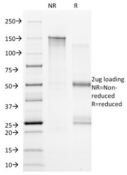
Biotium CD20(B9E9), CF568 conjugate, 0.1mg/mL
Recognizes a protein of 33-37 kDa, identified as CD20 (Workshop V; Code CD20.12). B9E9 recognizes extracellular domain of CD20. The epitope is similar to or identical to that recognized by other CD20 antibodies including Leu-16 and B1. This MAb can be used for immunophenotyping of leukemia and malignant cells, B lymphocyte detection in peripheral blood, B cell localization in tissues and B lymphocyte purification by immunosorbent methods. CD20 is a non-Ig differentiation antigen of B-cells and its expression is restricted to normal and neoplastic B-cells, being absent from all other leukocytes and tissues. CD20 is expressed by pre B-cells and persists during all stages of B-cell maturation but is lost upon terminal differentiation into plasma cells. Protein passes through the membrane 4 times with both ends in cytoplasm and exposes one short and one longer loop to the external environment. CD20 is not glycosylated in resting B cells and its cytoplasmic domains are differentia
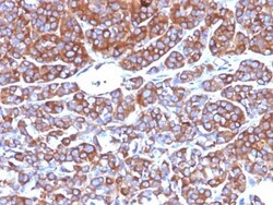
Biotium CD10(CB-CALLA), CF568 conjugate, 0.1mg/mL
CD10 (also known as Common Acute Lymphocytic Leukemia Antigen (CALLA)) is a type II transmembrane protein. It is a cell surface enzyme with neutral metalloendopeptidase activity, which inactivates a variety of biologically active peptides. CD10 is expressed on the cells of lymphoblastic, Burkitt's, and follicular germinal center lymphomas, and on cells from patients with chronic myelocytic leukemia (CML). It is also expressed on the surface of normal early lymphoid progenitor cells, immature B cells within adult bone marrow and germinal center B cells within lymphoid tissue.Primary antibodies are available purified, or with a selection of fluorescent CF Dyes and other labels. CF Dyes offer exceptional brightness and photostability. Note: Conjugates of blue fluorescent dyes like CF405S and CF405M are not recommended for detecting low abundance targets, because blue dyes have lower fluorescence and can give higher non-specific background than other dye colors.

Enzo Life Sciences 8-(4-Chlorophenylthio)guanosine 3',5'-cyclic Monophosphate . triethylammonium salt (5mg). CAS: 54364-02-2
Selective activator of cGMP-dependent protein kinase Iα, Iβ and type II as well as of cGMP-gated ion channels. Higher activation potential as compared to cGMP; excellent cell membrane permeability and phosphodiesterase stability. Preferred alternative to unsuitable analogs such as dibutyryl-cGMP. Alternative name: 8-pCPT-cGMP . TEA. Purity: ≥99% (HPLC). Appearance: White to off-white powder. Solubility: Soluble in water. Long Term Storage: -20°C.
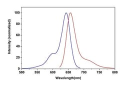
Abcam Phalloidin iFluor 633 Reagent
Phalloidin iFluor 633 Reagent
The product is subject to the following: Abcam Restricted Use Statement
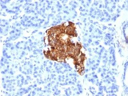
Biotium Insulin(E2-E3), CF488A conjugate, 0.1mg/mL
Recognizes a polypeptide which is identified as insulin, a 51-amino acid polypeptide composed of A and B chains connected through the C-peptide. Proinsulin, which has very little biological activity, is cleaved by proteases within its cell of origin into the insulin molecule and the C-terminal basic residue. Insulin enhances membrane transport of glucose, amino acids, and certain ions. It also promotes glycogen storage, formation of triglycerides, and synthesis of proteins and nucleic acids. Deficiency of insulin results in diabetes mellitus. The main storage site for insulin is the pancreatic islets. Antibodies to insulin are important as beta-cell and insulinoma marker.Primary antibodies are available purified, or with a selection of fluorescent CF Dyes and other labels. CF Dyes offer exceptional brightness and photostability. Note: Conjugates of blue fluorescent dyes like CF405S and CF405M are not recommended for detecting low abundance targets, because blue dyes have
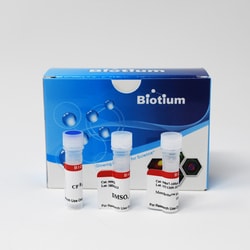
Biotium MemBrite™ Fix 640/660 Cell Surface Staining Kit, 500 Reactions
MemBrite™ Fix Cell Surface Staining Kits are designed for covalently staining the surface of live cells to conveniently visualize cell boundaries in immunofluorescence experiments. MemBrite™ Fix are new covalent cell surface stains that react irreversibly with cell surface proteins for staining that can withstand formaldehyde or alcohol fixation, and detergent permeabilization. Unlike lectins such as WGA, which bind specific targets that may vary between cell types, MemBrite™ Fix dyes react widely with cell surface proteins. MemBrite™ Fix staining is rapid and non-toxic to cells, and because MemBrite™ Fix dyes are highly water soluble, they stain cells much more evenly than traditional lipophilic membrane dyes like DiO, DiI, PKH, Vybrant, or CellMask™. This kit has reagents sufficient for 500 labeling reactions. The kit contains 5 vials of MemBrite™ Fix 640/660. Each vial is sufficient for 20 uL of 1000X dye after DMSO reconstitution.
Biotium CD86(BU63), CF488A conjugate, 0.1mg/mL
Recognizes a protein of 70 kDa, which is identified as CD86. CD86, along with CD80/B71, is an important accessory molecule in T cell co-stimulation via its interaction with CD28 and CD152/CTLA4, a cancer immunotherapy target. It is a type I transmembrane glycoprotein and a member of the immunoglobulin superfamily of cell surface receptors. It is expressed at high levels on resting peripheral monocytes and dendritic cells and at very low density on resting B and T lymphocytes. CD86 expression is rapidly upregulated by B cell specific stimuli with peak expression at 18 to 42 hours after stimulation. Since CD86 has rapid kinetics of induction, it is believed to be the major CD28 ligand expressed early in the immune response. It is also found on malignant Hodgkin and Reed Sternberg (HRS) cells in Hodgkin's disease.Primary antibodies are available purified, or with a selection of fluorescent CF Dyes and other labels. CF Dyes offer exceptional brightness and photostability. N

Medchemexpress LLC IL-17A Rat CHO 10ug | 10ug

The IL-17A protein has important heterodimerization and homodimerization activities and is involved in a variety of processes including responses to glucocorticoid stimulation and the regulation of cell death and transcription IL-17A is present in the cytoplasm and extracellular space and has been implicated in diseases such as pancreatitis inflammatory bowel disease renal disease and periodontal disease and may serve as a biomarker IL-17A Protein Rat (CHO) is the recombinant rat-derived IL-17A protein expressed by CHO with tag free
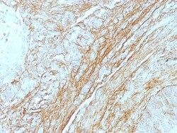
Biotium Tenascin(T2H5), CF568 conjugate, 0.1mg/mL
In Western blotting, this antibody reacts with two bands of ~MW of 210 kDa and 300 kDa, identified as two isoforms of Tenascin C. Specificity of this MAb is validated by sequential immunoprecipitation with a PAb against Tenascin C. Tenascin C is a multifunctional, disulfide-linkedhexameric extracellular matrix glycoprotein expressed in association with mesenchymal epithelial interactions during development and in the neo-vasculature and stroma of undifferentiated tumors. In adults, it is restricted to certain epithelial-stromal interfaces and increases markedly in hyper-proliferative diseases and in stroma of many neoplasms, including gliomas, breast, squamous and lung carcinomas.Primary antibodies are available purified, or with a selection of fluorescent CF Dyes and other labels. CF Dyes offer exceptional brightness and photostability. Note: Conjugates of blue fluorescent dyes like CF405S and CF405M are not recommended for detecting low abundance targets, because blue
Biotium FOXP3(FXP3/197), CF488A conjugate, 0.1mg/mL
Recognizes a protein of 47-55 kDa, which is identified as FOXP3. Its precise epitope is not known, but it has been mapped to the N-terminal portion of the protein. The FOX family of transcription factors is a large group of proteins that share a common DNA binding domain termed a winged-helix or forkhead domain. During early development, FOXP1 and FOXP2 are expressed abundantly in the lung, with lower levels of expression in neural, intestinal and cardiovascular tissues, where they act as transcription repressors. FOXP1 is widely expressed in adult tissues, while neoplastic cells often exhibit a dramatic change in expression level or localization of FOXP1. Mutations in FOXP3 gene cause IPEX, a fatal, X-linked inherited disorder characterized by immune dysregulation. The FOXP3 protein is essential for normal immune homeostasis. Specifically, FOXP3 represses transcription through a DNA binding forkhead domain, thereby regulating T cell activation.Primary antibodies are availabl
Biotium CD2(1E7E8.G4), CF568 conjugate, 0.1mg/mL
CD2 interacts through its amino-terminal domain with the extracellular domain of CD58 (also designated CD2 ligand) to mediate cell adhesion. CD2/CD58 binding can enhance antigen-specific T cell activation. CD2 is a transmembrane glycoprotein that is expressed on peripheral blood T lymphocytes, NK cells and thymocytes. CD58 is a heavily glycosylated protein with a broad tissue distribution in hematopoietic and other cells, including endothelium. Interaction between CD2 and its counter receptor LFA3 (CD58) on opposing cells optimizes immune system recognition, thereby facilitating communication between helper T lymphocytes and antigen-presenting cells, as well as between cytolytic effectors and target cells.Primary antibodies are available purified, or with a selection of fluorescent CF Dyes and other labels. CF Dyes offer exceptional brightness and photostability. Note: Conjugates of blue fluorescent dyes like CF405S and CF405M are not recommended for detecting low abunda
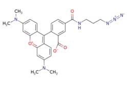
eMolecules 825651-66-9 | Medchem Express | TAMRA azide 5-isomer | 1mg | 784544163 | HY-151857 | 512.57 | C28H28N6O4
Medchem Express | TAMRA azide 5-isomer | 1mg | 784544163 | HY-151857 | 825651-66-9 | 512.570 | C28H28N6O4
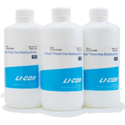
LICOR IRDye 800CW NHS Ester (250mg)
IRDye infrared dyes from LI-COR Biosciences are available for researchers developing applications using near-infrared (NIR) fluorescence. These unique dyes are used in a variety of NIR imaging applications including Western blotting, plate-based assays, protein arrays, tissue section imaging, and molecular activity measurements. Standard NHS ester chemistry is used to produce custom probes labeled with LI-COR IRDye infrared dyes. The NHS ester reactive group provides the functionality for labeling primary and secondary amines, such as lysine residues in proteins.